Hypertrophic Cardiomyopathy - C30 Auscultation Lesson with Recordings
Virtual Auscultation
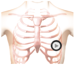
The patient's position is supine.
Lesson
An early peaking, harsh diamond shaped systolic murmur starts at the beginning of systole and ends well before the second heart sound. A fourth heart sound gallop is also present in diastole as you can readily see on the wave form tab. S1 is increased due to a hyperdynamic left ventricle. S2 is single. On the anatomy video you can see that the contraction of the left ventricle is strong and occurs in a reduced amount of time. Anatomically, the septal wall is very much thicker than the rest of the ventricle but this is not shown in the animation. The strong contraction of the left ventricle causes the anterior leaflet to be sucked into the ventricle, blocking the flow into the aorta and causing an aortic murmur. At the same time turbulent flow from the left ventricle to the left atrium causes a second murmur. Since the two murmurs occur at the same time you hear a single murmur. You can hear the difference between the two murmurs by moving the stethoscope head the aortic to the mitral valve area. First, you will hear the diamond shaped aortic murmur and later the rectangular pansystolic murmur.Waveform
Heart Sounds Video
Observing the animation, you can see that the contraction of the left ventricle is strong and occurs in a reduced amount of time. Anatomically, the septal wall is very much thicker than the rest of the ventricle but this is not shown in the animation.
Authors and Sources
Authors and Reviewers
-
Heart sounds by Dr. Jonathan Keroes, MD and David Lieberman, Developer, Virtual Cardiac Patient.
- Lung sounds by Diane Wrigley, PA
- Respiratory cases: William French
-
David Lieberman, Audio Engineering
-
Heart sounds mentorship by W. Proctor Harvey, MD
- Special thanks for the medical mentorship of Dr. Raymond Murphy
- Reviewed by Dr. Barbara Erickson, PhD, RN, CCRN.
-
Last Update: 12/11/2022
Sources
-
Heart and Lung Sounds Reference Library
Diane S. Wrigley
Publisher: PESI -
Impact Patient Care: Key Physical Assessment Strategies and the Underlying Pathophysiology
Diane S Wrigley & Rosale Lobo - Practical Clinical Skills: Lung Sounds
- Essential Lung Sounds
Diane S. Wrigley, PA-C
Published by MedEdu LLC - PESI Faculty - Diane S Wrigley
-
Case Profiles in Respiratory Care 3rd Ed, 2019
William A.French
Published by Delmar Cengage - Essential Lung Sounds
by William A. French
Published by Cengage Learning, 2011 - Understanding Lung Sounds
Steven Lehrer, MD
- Clinical Heart Disease
W Proctor Harvey, MD
Clinical Heart Disease
Laennec Publishing; 1st edition (January 1, 2009)