Myocarditis
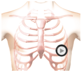
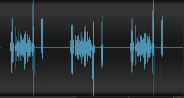
Here is a simulation of Myocarditis taken at the apex. 1. The first heart sound is softer than normal because of decreased function of the left ventricle. 2. The second heart sound is normal at the mitral area. 3. There is a third heart sound caused by the failure of the left ventricle. 4. A rectangular, medium-pitched murmur of mild mitral regurgitation is caused by the incomplete closure of the mitral valve leaflets. In the anatomy video you can see the enlarged left ventricle with decreased vigor of contraction. You can see the regurgitant turbulent flow from the left ventricle into the left atrium which is responsible for the murmur. Myocarditis is often the result of a viral infection of the myocardium.Auscultation Sounds
Position

The patient's position should be seated.
Listening Tips for Myocarditis
S1:SofterSystole:Rectangular, medium pitch murmur
Diastole:S3
Waveform (Phonocardiogram)
Observe Cardiac Animation for Myocarditis
Play the animation, taking note of the enlarged left ventricle with decreased vigor of contraction.
Observe the regurgitant turbulent flow from the left ventricle into the left atrium which is responsible for the murmur.
Authors and Sources
Authors and Reviewers
- EKG heart rhythm modules: Thomas O'Brien.
- EKG monitor simulation developer: Steve Collmann
-
12 Lead Course: Dr. Michael Mazzini, MD.
- Spanish language EKG: Breena R. Taira, MD, MPH
- Medical review: Dr. Jonathan Keroes, MD
-
Heart sounds and mentorship: W. Proctor Harvey, MD
- Medical review: Dr. Pedro Azevedo, MD, Cardiology
-
Last Update: 1/8/2023
Sources
-
Electrocardiography for Healthcare Professionals, 5th Edition
Kathryn Booth and Thomas O'Brien
ISBN10: 1260064778, ISBN13: 9781260064773
McGraw Hill, 2019 -
Rapid Interpretation of EKG's, Sixth Edition
Dale Dublin
Cover Publishing Company -
12 Lead EKG for Nurses: Simple Steps to Interpret Rhythms, Arrhythmias, Blocks, Hypertrophy, Infarcts, & Cardiac Drugs
Aaron Reed
Create Space Independent Publishing -
Heart Sounds and Murmurs: A Practical Guide with Audio CD-ROM 3rd Edition
Elsevier-Health Sciences Division
Barbara A. Erickson, PhD, RN, CCRN - Clinical Heart Disease
W Proctor Harvey, MD
Clinical Heart Disease
Laennec Publishing; 1st edition (January 1, 2009) -
The Virtual Cardiac Patient: A Multimedia Guide to Heart Sounds, Murmurs, EKG
Jonathan Keroes, David Lieberman
Publisher: Lippincott Williams & Wilkin)
ISBN-10: 0781784425; ISBN-13: 978-0781784429 - Project Semilla, UCLA Emergency Medicine, EKG Training Breena R. Taira, MD, MPH
Myocarditis | Lessons with Audio and Video | #115