Ventricular Septal Defect
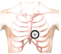
This is an example of ventricular septal defect as heard at the tricuspid position. Ventricular Septal Defect is a congenital condition associated with abnormal blood flow between the left ventricle and the right ventricle. During fetal development a wall develops creating a right and left ventricle. In a percentage of individuals a defect in the wall persists allowing blood flow from the left ventricle into the right ventricle. This condition is known as a ventricular septal defect. The first heart sound is normal. The second heart sound is unsplit. There is a third heart sound followed by a short diamond shaped diastolic murmur. A medium pitched murmur fills all of systole. In the anatomy video you see an enlarged right ventricle and an enlarged left atrium. You see turbulent blood flow from the left ventricle into the right ventricle through the up portion of the septum (the systolic murmur). There is further turbulent flow into the left ventricle from the left atrium causing the diastolic murmur. This is caused by VSD induced increased blood flow across the mitral valve.Auscultation Sounds
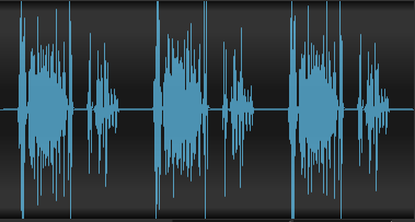
Patient Recording of Ventricular Septal Defect
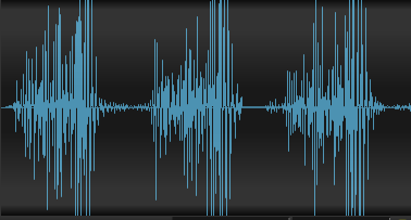
Patient Recording - Half Speed
Position
The patient's position should be supine.
Listening Tips for Ventricular Septal Defect
Diastole:S3 in patient recordings. Simulated sound includes a brief murmurWaveform (Phonocardiogram)
Observe Cardiac Animation for Ventricular Septal Defect
Observe an enlarged right ventricle and an enlarged left atrium in the cardiac animation.
Authors and Sources
Authors and Reviewers
- EKG heart rhythm modules: Thomas O'Brien.
- EKG monitor simulation developer: Steve Collmann
-
12 Lead Course: Dr. Michael Mazzini, MD.
- Spanish language EKG: Breena R. Taira, MD, MPH
- Medical review: Dr. Jonathan Keroes, MD
-
Heart sounds and mentorship: W. Proctor Harvey, MD
- Medical review: Dr. Pedro Azevedo, MD, Cardiology
-
Last Update: 1/8/2023
Sources
-
Electrocardiography for Healthcare Professionals, 5th Edition
Kathryn Booth and Thomas O'Brien
ISBN10: 1260064778, ISBN13: 9781260064773
McGraw Hill, 2019 -
Rapid Interpretation of EKG's, Sixth Edition
Dale Dublin
Cover Publishing Company -
12 Lead EKG for Nurses: Simple Steps to Interpret Rhythms, Arrhythmias, Blocks, Hypertrophy, Infarcts, & Cardiac Drugs
Aaron Reed
Create Space Independent Publishing -
Heart Sounds and Murmurs: A Practical Guide with Audio CD-ROM 3rd Edition
Elsevier-Health Sciences Division
Barbara A. Erickson, PhD, RN, CCRN - Clinical Heart Disease
W Proctor Harvey, MD
Clinical Heart Disease
Laennec Publishing; 1st edition (January 1, 2009) -
The Virtual Cardiac Patient: A Multimedia Guide to Heart Sounds, Murmurs, EKG
Jonathan Keroes, David Lieberman
Publisher: Lippincott Williams & Wilkin)
ISBN-10: 0781784425; ISBN-13: 978-0781784429 - Project Semilla, UCLA Emergency Medicine, EKG Training Breena R. Taira, MD, MPH
Ventricular Septal Defect | Lessons with Audio and Video | #111